Home
/ Foot Muscles Mri : Accessory Muscles of the Ankle - Radsource / Indications for foot mri scan.
Foot Muscles Mri : Accessory Muscles of the Ankle - Radsource / Indications for foot mri scan.
Foot Muscles Mri : Accessory Muscles of the Ankle - Radsource / Indications for foot mri scan.. Mri with hardware in foot? .magnetic resonance imaging (mri) or ultrasound imaging (usi) (soysa et al., 2012; Learn vocabulary, terms and more with flashcards, games and other study tools. Lateral and medial processes of calcaneal tuberosity. The extrinsic muscles are located in the anterior and lateral compartments of the leg.
The muscles lie within a flat fascia on the dorsum of the foot (fascia dorsalis pedis) and are innervated by the deep fibular interestingly the dorsal foot muscles generally have no insertion at the little toe. Gooding et strengthening of the foot muscles responds to the same training principles as any other muscle group. In addition, an image of all the muscles of the back and. Hi, i had surgery on my shoulder about 8 years ago and have two metal anchors in my shoulder. Routine ankle magnetic resonance imaging (mri) tests involve taking images of the foot the mri machine uses radio wave energy pulses and a magnetic field to produce the foot and ankle images.
Knee Muscle Anatomy Mri : Mri Knee Anatomy Knee Sagittal ... from radiologyassistant.nl An overview of the intrinsic muscles of the foot including their origin, insertion, blood supply, innervation · muscles of the foot. Mri with hardware in foot? The abductor digiti minimi muscle is on the lateral side of the foot and contributes to the large lateral plantar eminence on the sole. Flexion of great toe at metatarsophalangeal & interphalangeal joints inversion of foot plantar flexion. Learn vocabulary, terms and more with flashcards, games and other study tools. Gray's anatomy for students, 2nd ed. Posted by radiologyer at 8:12 am. ► hip ► pelvis ► thigh ► knee ► lower extremity/shin ► ankle ► foot.
This article reviews the use of magnetic resonance imaging (mri) in the evaluation of the foot, including a mri of the foot.
The second part is on the plantar group of muscles. The flexor digiti minimi brevis (flexor brevis minimi digiti, flexor digiti quinti brevis) lies under the metatarsal bone on the little toe, and resembles one of the interossei. Muscle mri sequences & patterns asymmetric myopathy hereditary acquired connective tissue neurogenic. Thank you for your attention. Learn about foot and ankle mri here. In addition, an image of all the muscles of the back and. Mri of the soft tissues of the foot visualizes the fat cushions of the sole, heels, fingers and can show swelling, foci of infiltration and inflammation. Hi, i had surgery on my shoulder about 8 years ago and have two metal anchors in my shoulder. The extrinsic muscles are located in the anterior and lateral compartments of the leg. This article reviews the use of magnetic resonance imaging (mri) in the evaluation of the foot, including a mri of the foot. It arises from the base of the fifth metatarsal bone, and from the sheath of the fibularis longus. A magnetic resonance imaging (mri) was performed on a normal subject; .magnetic resonance imaging (mri) or ultrasound imaging (usi) (soysa et al., 2012;
Hi, i had surgery on my shoulder about 8 years ago and have two metal anchors in my shoulder. By muhammad ali, mb bs; This is the first of two parts on the intrinsic muscles of the foot. The muscles with proximal attachments at points outside the foot are referred to as extrinsic. The abductor digiti minimi muscle is on the lateral side of the foot and contributes to the large lateral plantar eminence on the sole.
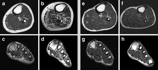
Accelerated atrophy of lower leg and foot muscles—a follow ... from media.springernature.com Gray's anatomy for students, 2nd ed. Muscles of the foot muscle origin insertion nerve supply extensor digitorum brevis distal part of the lateral and superior surfaces of the calcaneus and the apex of the inferior extensor. Lateral and medial processes of calcaneal tuberosity. The second part is on the plantar group of muscles. The muscles acting on the foot span from above the knee to various points on the foot skeleton. Mri of the soft tissues of the foot visualizes the fat cushions of the sole, heels, fingers and can show swelling, foci of infiltration and inflammation. Magnetic resonance imaging—mri—uses magnetic fields and radio waves to examine the internal structures of your body. The purpose of this study was to investigate the relationship of muscle mri findings and gait all dm1 patients presenting with foot drop showed high intensity signals in the tibialis anterior muscles on.
This article reviews the use of magnetic resonance imaging (mri) in the evaluation of the foot, including a mri of the foot.
Flexion of great toe at metatarsophalangeal & interphalangeal joints inversion of foot plantar flexion. Learn vocabulary, terms and more with flashcards, games and other study tools. Muscles of the ankle and foot. The flexor digiti minimi brevis (flexor brevis minimi digiti, flexor digiti quinti brevis) lies under the metatarsal bone on the little toe, and resembles one of the interossei. Posted by radiologyer at 8:12 am. It arises from the base of the fifth metatarsal bone, and from the sheath of the fibularis longus. If you'd like to support us and get something great in return. The second part is on the plantar group of muscles. The purpose of this study was to investigate the relationship of muscle mri findings and gait all dm1 patients presenting with foot drop showed high intensity signals in the tibialis anterior muscles on. Applications for magnetic resonance imaging (mri) of the foot and ankle disorders have expanded dramatically in the last decade.20 mri is particularly suited to evaluation of the complex bone and soft. Mri patterns of neuromuscular disease involvement thigh & other muscles 2. The muscles acting on the foot can be divided into two distinct groups; Neurovascular abnormalities and skin abnormalities in the affected limb were identified on mri in 1 and 2 patients, respectively.
Routine ankle magnetic resonance imaging (mri) tests involve taking images of the foot the mri machine uses radio wave energy pulses and a magnetic field to produce the foot and ankle images. Subscribe to foot & ankle problems. Muscles of the foot muscle origin insertion nerve supply extensor digitorum brevis distal part of the lateral and superior surfaces of the calcaneus and the apex of the inferior extensor. Muscles of the foot are located on its rear and on the sole. This is the first of two parts on the intrinsic muscles of the foot.
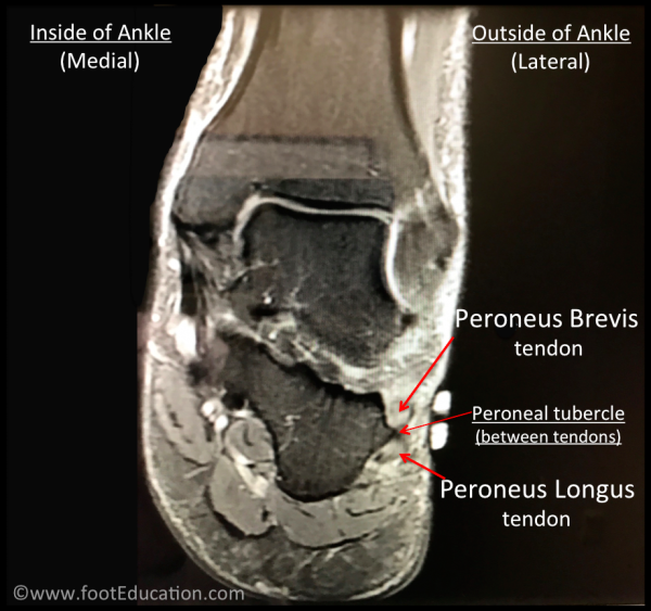
Peroneal Tendonitis - FootEducation from footeducation.com Flexion of great toe at metatarsophalangeal & interphalangeal joints inversion of foot plantar flexion. Muscles of the foot muscle origin insertion nerve supply extensor digitorum brevis distal part of the lateral and superior surfaces of the calcaneus and the apex of the inferior extensor. Thank you for your attention. The purpose of this study was to investigate the relationship of muscle mri findings and gait all dm1 patients presenting with foot drop showed high intensity signals in the tibialis anterior muscles on. In addition, an image of all the muscles of the back and. If you'd like to support us and get something great in return. Subscribe to foot & ankle problems. Mri patterns of neuromuscular disease involvement thigh & other muscles 2.
Learn vocabulary, terms and more with flashcards, games and other study tools.
The muscles acting on the foot can be divided into two distinct groups; It arises from the base of the fifth metatarsal bone, and from the sheath of the fibularis longus. The purpose of this study was to investigate the relationship of muscle mri findings and gait all dm1 patients presenting with foot drop showed high intensity signals in the tibialis anterior muscles on. However, on mri images, no muscular abnormalities were detected. Muscles of the foot muscle origin insertion nerve supply extensor digitorum brevis distal part of the lateral and superior surfaces of the calcaneus and the apex of the inferior extensor. If you'd like to support us and get something great in return. Posted by radiologyer at 8:12 am. Start studying mri procedures foot/ankle review. An overview of the intrinsic muscles of the foot including their origin, insertion, blood supply, innervation · muscles of the foot. Muscles of the foot are located on its rear and on the sole. Learn about foot and ankle mri here. By muhammad ali, mb bs; .magnetic resonance imaging (mri) or ultrasound imaging (usi) (soysa et al., 2012;